Best-in-class equipment & specialists keeping your vision perfect as the Eagle!
Our Location
Shop M8, Orchid Shopping Mall, Lekki, Lagos.

Call Us
08030482260
Your Cart Details
Comprehensive Eye Tests
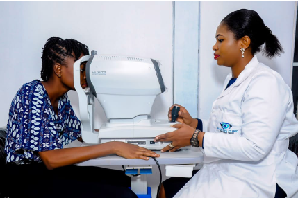
Raint Eye Clinic we deliver a comprehensive service. Each vision examination is individualised and takes 45 minutes.
We believe a longer assessment gives us time to understand your visual needs, so we can help you reach the best visual solution. We routinely diagnose and manage conditions which can affect the eye such as glaucoma, cataract, retinal disease, diabetes, or squint. Treatments are available for many of these conditions especially if they are detected early. Our practice has a number of specialised instruments which help assess and monitor eye disease.
Do I need a vision examination?
We recommend comprehensive vision examinations every 2-3 years. Eye diseases such as glaucoma, have no symptoms at very early stages but can be picked up at routine examinations. After your examination your Optometrist will recommend a recall appropriate to your eyes and vision. This is likely to be at least every two years, although we often see those with eye disease, or a family history of disease, more often.
What are the most common visual symptoms?
Most visual problems have associated symptoms. Some of the more common symptoms include:
- Blur in the distance, at near or both
- Headaches when reading
- Rubbing eyes repeatedly
- Dislike or avoidance of close work
- Holding head at an angle when looking at an object
- Shutting or covering one eye to focus
- Excessive blinking
- Holding books closely
- Using a finger as a place mark when reading
- Sitting very close to objects such as the television
- Developing red eyes
- Discomfort within or around the eyes
What happens during a comprehensive vision examination?
During your examination, our Optometrists use a series of tests to measure the strength of prescription lenses, if necessary, for clear comfortable vision. They will assess the health of both the outside tissues of your eyes and eyelids and also examine the tissues inside your eyes. This will be followed by a customised management plan for your care.
Possible outcomes include:
- Spectacles
- Contact Lenses
- Eye Surgery
- Low Visual Aids
- Visual Therapy
- Dry eye management
- Myopia Control
- Medical Eye treatments
- Tinted & Coloured Lenses
- Sports Vision Enhancement
- Further in-depth assessment and monitoring
- Referral to other Health Professional
The following is a list of the steps we commonly take during a vision examination. Every examination is individualised, so we may spend more time on one area depending on your circumstances and eye history.
History
We discuss your family history of eye disease, your current spectacles, contact lenses, current medication(s), your general health, and individual eye history such as treatments or injuries.
Acuity testing
Acuity testing tests how sharp your vision is. Your vision will be examined at both distance and up close. You will be asked to read an eye chart with each eye. The smaller the letters that can be distinguished, the better your visual acuity. Your Optometrist will carefully test eye muscle balance to see how easily the eyes move and focus individually as well as together.
Refraction
Refraction is the process by which the Optometrist determines your ‘prescription’. This is the correction factor required to help you see at a certain distance.
Microscope Examination
A non-invasive examination of the eye both inside and out is crucial in the diagnosis of eye disease. The cornea is regarded as the clear “window” of the eye. A ‘slit lamp’ microscope is used to examine the cornea. This microscopic examination of the cornea allows your Optometrist to check for irregularities. The test is external and completely painless. Other structures examined include your eyelids, lashes, tear film, conjunctiva, ocular lens, anterior chamber angles, punctum and tear drainage system and oil producing glands.
Glaucoma Pressure Test
Tonometry is a measure of the pressure in your eye, related to the production and outflow of fluid. If the pressure in the eye is high, this can cause damage to the optic nerve and result in glaucoma. Unless your eye pressure is extremely high, an increase in this is usually painless and therefore comes without symptoms.
Keratometry / Topography
Using keratometry or topography your Optometrist measures the curvature of the corneal surface. This is particularly important for those interested in contact lenses; we can find the best fit for you when we have a measurement of your corneal shape.
Ophthalmoscopy
Using an ophthalmoscope, a non-invasive, hand held instrument, your Optometrist can conduct an examination of the inside of the eye. The ophthalmoscope has a bright light, which illuminates and magnifies the interior of the eye. Cataracts, retinal problems, damaged blood vessels can be found in this examination. Your health can also tell us a lot about your general health. For those who have diabetes or previously undetected high blood pressure, we often see characteristic changes to the blood vessels.
Pupil Dilation
Pupil dilation means temporarily enlarging the pupil. This is done with eye drops. Dilation allows a more complete internal view of the eye and is often used when vision is reduced or in elderly individuals. The pupil of the eye returns to normal when the drops wear off after a few hours. The time for these to wear off varies from person to person, so we recommend you have someone drive you home. Wearing sunglasses after pupil dilation relieves glare.
Retinal Photography
The retinal camera takes an image of the posterior ocular surface. This provides us with detailed colour digital images of the pigment layer, major blood vessels, optic nerve head and posterior structures for future reference and comparison.
Ocular Coherence Tomography (OCT)
Ocular coherence tomography is a new and modern technology within Optometry. The OCT uses light to deliver high definition images of your eye and its structures. These measurements are recorded in microns – a micron being one 1000th of a millimetre. An OCT scan is considered the gold-standard of care for monitoring or the early diagnosis of conditions such as:
- Glaucoma
- Macular degeneration
- Narrow anterior chamber angles
- Keratoconus or corneal disease
- Macular holes
Meet Our Optometrists
We Have Best Professional Team To Care Your Eyes